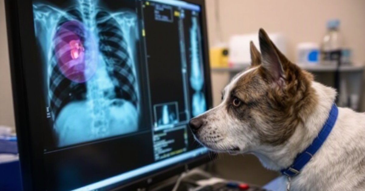
When Your Pet Swallows Something, the Scan Matters as Much as the Speed
Gut blockages in dogs and cats — caused by swallowed objects that get stuck in the digestive tract — are one of the most urgent emergencies vets face. A new study suggests that ultrasound may catch many of these blockages that a standard X-ray would miss, and that using both imaging tools together improves the odds of an accurate diagnosis. For pet owners, that finding has a clear take-home message: if your dog or cat swallowed something and the X-ray looks normal, that doesn’t necessarily mean everything is fine.
Researchers reviewed 20 years of imaging records from dogs and cats treated for gastrointestinal foreign body obstructions — objects lodged in the stomach or intestines. Their conclusion: ultrasound frequently detected the obstruction directly, and a clean X-ray alone was not always enough to rule one out.
Why X-Rays Don’t Always Tell the Whole Story
X-rays — also called radiographs — have long been the first step vets take when a pet is suspected of swallowing something dangerous. They’re fast, widely available, and excellent at showing dense objects like bones, coins, or metal screws. But many swallowed objects are made of softer materials: fabric, rubber, plastic, corn cobs, toys. These items don’t show up clearly on X-rays. They can blend right in with the tissue around them, making the image look deceptively normal.
Ultrasound works differently. Instead of using radiation to take a snapshot of the body’s interior, it sends high-frequency sound waves through the body and maps how those waves bounce back — a bit like sonar on a submarine. Because it picks up soft tissues and fluid in real time, ultrasound can often directly show a foreign object that an X-ray would miss entirely.
The gap between what these two tools can detect is exactly what this study set out to measure.
What the Researchers Looked At
This was a retrospective study — meaning researchers looked back through patient records rather than conducting a new experiment. The team analyzed 20 years of imaging data from animals referred with suspected gastrointestinal foreign body obstructions.
The reviewed population included:
- 411 dogs and 78 cats referred with suspected obstruction
- 106 dogs and 29 cats confirmed to have a true obstruction
For each case, the researchers examined the imaging results — which tests were used, what each test showed, and how the findings lined up with the confirmed diagnosis. The goal was to understand how well X-rays and ultrasound each performed, and whether combining them made a meaningful difference.
What the Study Found
Ultrasound Frequently Spotted What X-Rays Could Miss
The study found that ultrasonography frequently directly visualized the foreign body — meaning the blocked object could actually be seen on the scan, not just inferred from indirect signs like gas buildup or a stretched bowel. This made ultrasound a strong primary imaging tool for this type of emergency.
That’s a meaningful distinction. An X-ray might suggest something is wrong by showing abnormal gas patterns or a distended loop of intestine — clues that something is off. But when ultrasound directly shows the foreign object itself, the vet doesn’t have to guess: the blockage is visible, its location is clearer, and the next step in treatment is easier to plan.
Combining Both Tools Improved Accuracy
The researchers also found that using both radiography (X-ray) and ultrasound together improved diagnostic performance compared to either tool alone. The two methods are complementary: X-rays capture the big picture quickly and can reveal some objects immediately, while ultrasound fills in the gaps for soft or oddly shaped foreign material.
Think of it like checking a room with two different kinds of lighting. A regular overhead light (X-ray) floods the space and shows you most things at once. A UV light (ultrasound) picks up stains, fibers, and fine details the overhead missed. Using both gives you the clearest picture of what’s actually there.
A Clear X-Ray Isn’t the Same as a Safe Pet
One of the study’s most practical findings: a normal or equivocal X-ray — one that looks fine or shows inconclusive results — does not reliably exclude a gastrointestinal obstruction. This is especially true in young animals with persistent vomiting or abdominal pain. In other words, if your puppy or kitten has been vomiting repeatedly and the X-ray didn’t show a problem, the possibility of a blockage may not be off the table.
What This Means If Your Pet Swallows Something
Don’t Wait for Multiple Symptoms to Pile Up
Gastrointestinal blockages are time-sensitive. The longer a foreign object sits in the digestive tract, the greater the risk of serious complications — including tissue damage or rupture. If your pet ate something they shouldn’t have, early imaging — even before symptoms are severe — gives the best chance of catching a problem while it’s still manageable.
This study suggests that ultrasound should be part of that early workup, not just a fallback when the X-ray is inconclusive.
Ask About Imaging Options at the Clinic
Not every vet clinic has an ultrasound machine, and not every technician has the same level of experience with abdominal ultrasound. If your vet’s X-ray comes back normal but your pet is still acting sick, it’s reasonable to ask whether an ultrasound is available, or whether a referral to an emergency or specialty clinic with better imaging resources makes sense.
You’re not second-guessing your vet — you’re being an informed advocate for your pet.
Signs That Should Send You to the Vet Right Away
If you think your dog or cat swallowed a non-food object, head to a vet promptly — don’t wait to see if it “passes.” Seek urgent care if your pet shows any of the following:
- Repeated vomiting, especially if it starts suddenly
- Straining without producing a bowel movement
- A swollen, tight, or painful-looking belly
- Loss of appetite paired with unusual lethargy
- Drooling or pawing at the mouth (more common in cats with throat obstructions)
- Known ingestion of something dangerous, like a corn cob, toy part, or piece of fabric
Young pets — puppies and kittens — are especially prone to swallowing objects and tend to deteriorate faster when a blockage is present, making early evaluation even more important.
Study Limitations
Because this was a retrospective study, the researchers were working with historical records rather than running a controlled experiment. That means findings reflect associations — patterns observed over 20 years — rather than a head-to-head test under identical conditions. The cases reviewed may not capture every scenario a vet might encounter, and variations in equipment quality, technician experience, and patient presentation across the 20-year window could affect the results.
As with any single study, these findings add to the evidence base rather than rewriting the book on how blockages should be diagnosed. Future prospective studies — ones that follow animals in real time under controlled conditions — would strengthen the conclusions further.
The Bottom Line
A 20-year look at imaging data from 106 dogs and 29 cats confirmed with gastrointestinal foreign body obstructions — out of 411 dogs and 78 cats referred with suspected obstruction — suggests that ultrasound is a powerful tool for diagnosing these emergencies, often directly showing blocked objects that X-rays can’t detect. Combining both imaging methods appears to improve diagnostic accuracy, and a normal X-ray alone should not be taken as reassurance when clinical signs point to a potential blockage.
If your pet swallows something and the X-ray is inconclusive, ask about ultrasound — and if you’re not getting answers, don’t hesitate to seek a second opinion at an emergency or specialty clinic. Speed and the right tools can make all the difference.
This article summarizes peer-reviewed research for educational purposes. Always consult with your veterinarian for personalized advice about your pet’s health and behavior.