Thermal Imaging in Dogs: Why Coat Type Changes the Picture
Dog thermal imaging results are significantly shaped by your dog’s coat length and the body region being scanned, according to a new study in Frontiers in Veterinary Science. Researchers measured surface temperatures in 33 healthy dogs and found that both coat length and body region were strong predictors of what the heat-sensing camera picked up — meaning a fluffy Samoyed and a sleek Whippet can look very different on a thermal scan even when both are perfectly healthy. That finding matters to any dog owner whose pet undergoes this kind of imaging for pain, injury, or rehab monitoring.
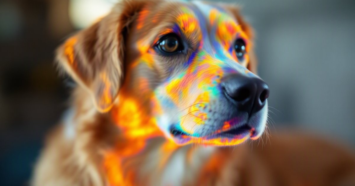
Thermal imaging, also called infrared thermography, is a non-invasive tool that detects heat coming off the skin’s surface. Hotter areas often signal inflammation or irritation; cooler areas can suggest reduced blood flow or nerve involvement. The technology is appealing because it requires no needles, no sedation, and no radiation. But for it to be useful, the person reading the scan needs to know what’s normal — and this study shows that “normal” varies quite a bit depending on the dog in front of the camera.
Why Coat Length Complicates Heat Readings
Think of a dog’s fur as a natural blanket. A thick coat traps heat close to the skin, which can either make a surface reading look warmer or insulate the skin temperature from the outside air, depending on how heat escapes through the fur. A short-haired dog has much less insulation between its skin and the camera, so the reading more directly reflects what’s happening in the tissue beneath.
Before this study, veterinary guidelines for thermal imaging didn’t fully account for how much coat length could shift a reading on its own — without any injury, inflammation, or disease present. That gap is what this research set out to address.
What the Study Involved
The researchers enrolled 33 healthy, client-owned adult dogs and used an infrared camera to record surface temperatures at multiple locations across the body. Importantly, all dogs were healthy, which allowed the team to establish clean baseline data — a picture of what normal thermal readings look like across different coat types and body areas.
Key details of the study:
- 33 healthy adult dogs owned by clients — a real-world sample, not lab animals
- Temperatures were measured at multiple body regions
- Coat length was recorded and analyzed as a variable
- Left-right comparisons were performed — comparing the same spot on both sides of the body
What the Researchers Found
Coat and Body Region Both Shift the Numbers
The clearest finding: coat length and body region each significantly influenced the temperature readings the camera captured. In plain terms, a long-coated dog will look different on a thermal scan than a short-coated dog in the same body area, even when both are healthy. Similarly, the same dog’s shoulder might read at a different temperature than its lower back — simply because of differences in muscle depth, blood vessel location, and fat coverage in those areas.
This doesn’t mean thermal imaging is unreliable. It means the results need context. Interpreting a scan without knowing the dog’s coat type — or without accounting for which body region is being measured — could lead to a misleading read.
Left-Right Symmetry Was Preserved
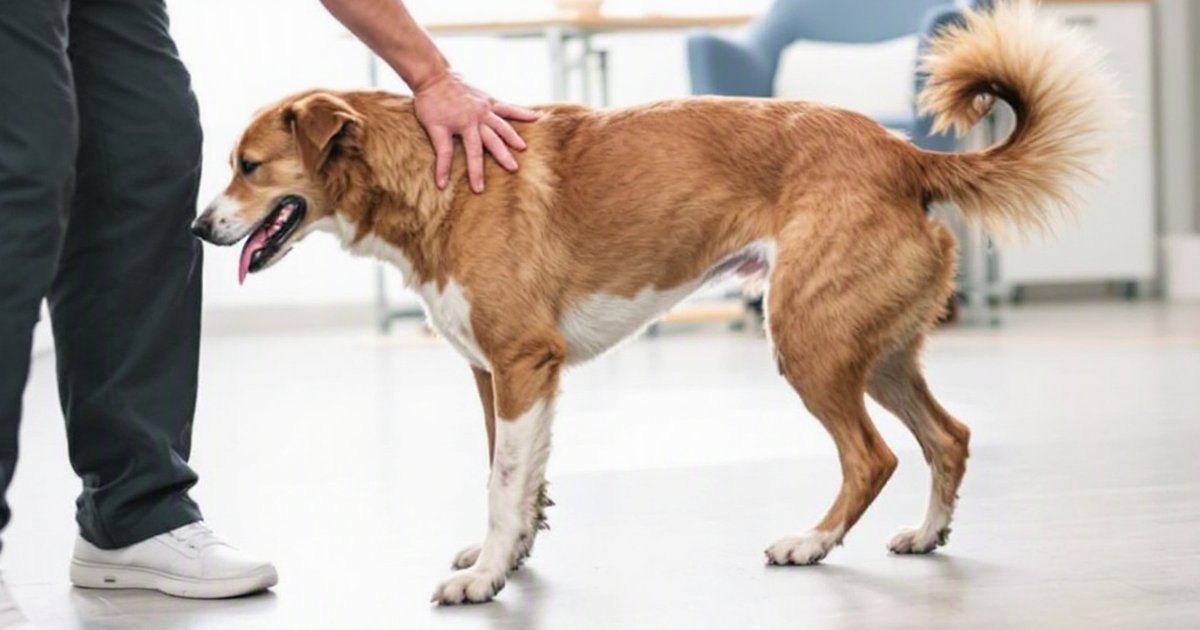
The encouraging finding: when comparing the same body spot on the left side versus the right side of the same dog, temperatures were generally symmetrical. This is important because side-to-side comparison is exactly how many vets and canine rehabilitation therapists use thermal imaging in practice. If the left shoulder looks significantly hotter than the right shoulder, that asymmetry is the clinical signal — it suggests something is different on one side.
The fact that left-right symmetry held up across dogs with different coat lengths suggests this comparison approach can still work reliably, as long as coat and body region are considered when drawing conclusions.
How This Research Applies to Your Dog
Coat Type Is Clinically Relevant Context
If your dog undergoes thermal imaging — for example, to look into back pain, monitor recovery after surgery, or screen for early joint inflammation — your dog’s coat type is relevant information for whoever reads the scan. A thick double coat in a Bernese Mountain Dog requires a different baseline than a short, single-layer coat in a Boxer.
This doesn’t mean you need to do anything special before an appointment. But it’s worth knowing that the professional interpreting the results should be applying coat-specific context, not a one-size-fits-all threshold.
Questions Worth Asking Your Vet or Rehab Therapist
If thermal imaging is offered as part of your dog’s care, consider asking:
- Has coat length been accounted for in the interpretation?
- Are you comparing the left and right sides of the body?
- Was any fur clipped before the scan? (Some clinics clip small areas for a cleaner skin reading.)
These aren’t gotcha questions — they’re the kind of specifics that help you understand whether the results you’re being shown are being read in context.
When Thermal Imaging Is Most Often Used
Thermal scanning tends to come up in these situations:
- Canine rehabilitation programs for dogs recovering from surgery or injury
- Assessing back or spinal pain, especially in breeds prone to disc problems (like Dachshunds and Corgis)
- Monitoring joint inflammation in dogs with arthritis
- Pre- and post-exercise screening in canine sport or working dogs
If your dog has any of these conditions or is going through a rehab program, it’s reasonable to ask whether thermal imaging is available and what training the person reading the results has had.
Study Limitations to Keep in Mind
This was a small observational study — just 33 dogs — and all subjects were healthy. That’s the right design for building baseline norms, but it doesn’t tell us how coat-related variation plays out in dogs that already have injuries or illness, where temperature signals may be harder to separate from background variation. The study also didn’t test whether adjusting for coat type actually improved diagnostic accuracy in clinical cases — that’s the important follow-up question. Larger studies across a wider range of breeds, ages, and health conditions will be needed to turn these findings into standardized clinical guidelines.
The Bottom Line
Thermal imaging in dogs is a promising non-invasive diagnostic tool — but coat length and body region significantly shape what the camera sees. A 33-dog prospective study found these two factors are strong predictors of surface temperature readings, meaning results can’t be interpreted the same way across all dogs. The good news is that left-right symmetry was generally preserved, so comparing both sides of the body remains a valid approach. If your dog is being assessed with thermal imaging, make sure the person reading the results is accounting for coat type and body region — not just flagging any temperature difference without that context.
This article summarizes peer-reviewed research for educational purposes. Always consult with your veterinarian for personalized advice about your pet’s health and behavior.