Where Vets Point the Ultrasound Wand Could Help Dogs in a Breathing Emergency
When a dog suddenly struggles to breathe after a car accident or chest injury, every second counts — and how vets use ultrasound to check for a collapsed lung may be more nuanced than it looks. A new pilot study published in Frontiers in Veterinary Science suggests that the exact placement of the ultrasound probe on a dog’s chest may affect how accurately a collapsed lung is identified in an emergency. The study compared different scanning locations and visual signs on the ultrasound screen to help emergency clinicians figure out the best approach — potentially making this quick, bedside tool even more reliable for dogs in crisis.
This kind of research is still early-stage (it used cadaver models rather than live patients), but the implications are real: even small improvements in how vets scan an injured dog’s chest could make a life-saving difference in the ER.
What a Collapsed Lung Actually Means for Your Dog
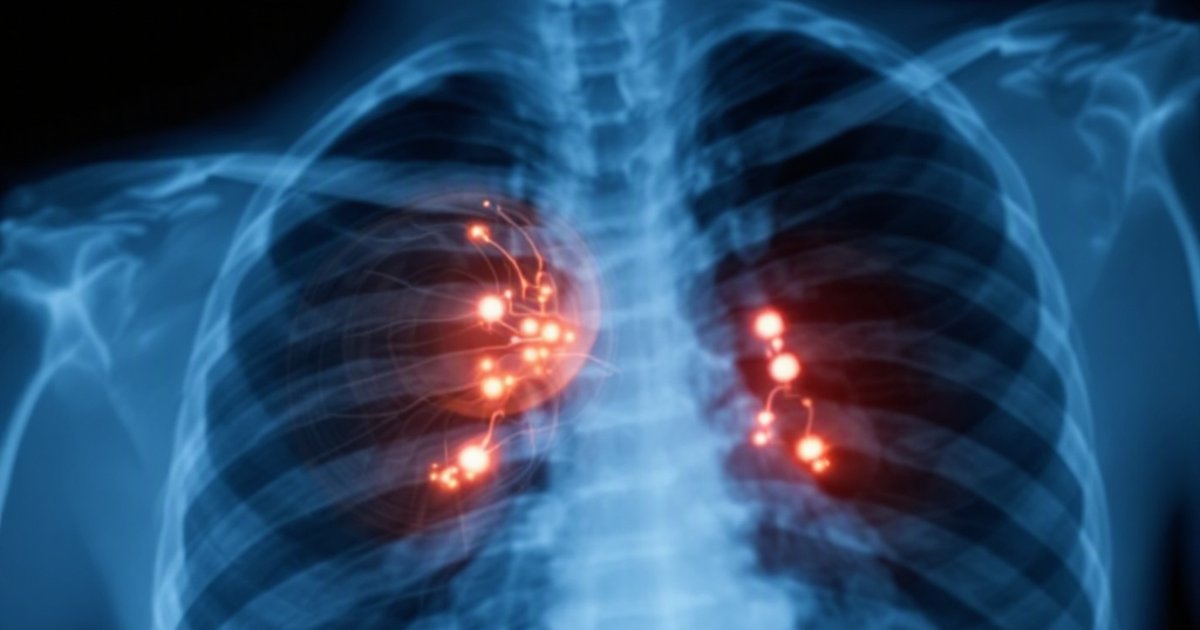
A collapsed lung, known medically as a pneumothorax (pronounced “new-mo-THOR-ax”), happens when air leaks into the space around the lung. Normally, the chest cavity is sealed, and the lung stays inflated like a balloon in a tight container. But if the chest wall is punctured — say, after a car accident, a fall, or a bite wound — air sneaks into that space and pushes on the lung from the outside. The lung can’t expand properly, and your dog can’t get enough oxygen.
Signs of a possible collapsed lung include:
- Sudden, labored breathing
- Rapid or shallow breaths
- Open-mouth breathing (unusual outside of panting)
- Blue or gray color on the gums
- Reluctance to move or a hunched posture
This is a veterinary emergency. If your dog shows any of these signs after a trauma, head straight to a 24-hour emergency vet — don’t wait for a regular appointment.
How Vets Currently Check for a Collapsed Lung
In an emergency, vets have a few tools for checking for a pneumothorax. X-rays are one option, but they take time and require the dog to stay still — tough when the animal is in distress. This is where point-of-care ultrasound (POCUS) comes in. Think of it as a handheld “look inside” tool: a vet presses the probe against the chest, and a live image appears on a screen within seconds. No waiting room, no radiation, no sedation required.
A healthy lung has a characteristic shimmer on ultrasound called a “glide sign” — that’s the lung wall gently sliding against the chest wall with each breath. When there’s a collapsed lung, that sliding motion disappears. The challenge is that the chest is a complex, curved structure, and where you put the probe matters — a lot. Place it in the wrong spot, and you might miss the very sign you’re looking for.
What the Researchers Studied
This pilot study used canine cadavers — dogs donated for research after passing away — to examine how different probe placements on the chest wall affect the ability to detect a pneumothorax. By deliberately introducing air into the chest cavity to simulate a collapsed lung, the team could test which scanning positions and which visual signs on the ultrasound screen were most useful.
Using cadavers rather than living dogs allowed researchers to control the experiment precisely — introducing a known amount of air and comparing what different scanning locations showed. It’s a standard approach for early diagnostic research, since it removes variables like movement, uneven breathing, or secondary injuries that might cloud the picture.
What the Study Found
Probe Placement Appears to Influence What You See
The study found that not all scanning positions on the chest wall are equally reliable for spotting a collapsed lung. Some locations may give emergency clinicians a clearer, more consistent view of the tell-tale loss of lung sliding — the main ultrasound sign that flags a pneumothorax.
The research also helped identify which visual patterns on the ultrasound screen are likely to be the most informative. In other words, it’s not just about knowing that the lung isn’t sliding — it’s about knowing where to look to catch that finding most reliably and quickly.
A Step Toward Smarter Emergency Scanning Protocols
By comparing multiple probe positions, the study helped narrow down a more systematic approach for dogs suspected of having a collapsed lung. Standardization matters enormously in emergency medicine. When a vet is working with an injured, frightened dog and needs answers fast, having a clear protocol for where to place the probe can reduce missed diagnoses and speed up treatment decisions.
What This Means If Your Dog Ever Needs Emergency Care
Faster, More Accurate Diagnosis Is the Goal
You may never think about ultrasound probe placement when rushing your dog to the emergency vet after an accident. But behind the scenes, this kind of detail separates a fast, confident diagnosis from an uncertain one. Research like this helps emergency vets build better scanning protocols — leading to fewer missed cases and quicker treatment.
For you as a pet owner, the practical takeaway is this: emergency ultrasound for chest injuries in dogs is a well-established, valuable tool — and the science behind it is actively being refined to make it even more reliable.
Signs That Mean Go Now
If your dog has been in any trauma — hit by a car, fallen from a height, bitten on the chest — watch closely for breathing changes in the hours that follow. A collapsed lung can sometimes develop or worsen gradually.
Don’t wait to see if it improves on its own. Call an emergency vet immediately if your dog:
- Breathes much faster or harder than normal after an injury
- Takes short, shallow breaths or appears to strain with each breath
- Has pale, blue, or grayish gums
- Becomes unusually quiet and reluctant to move
Study Limitations to Keep in Mind
Because this was a pilot study using cadavers, the findings need to be confirmed in living dogs before they can change standard clinical practice. Cadavers don’t breathe, and live dogs may have chest wall movement, muscle tension, or secondary injuries that affect the ultrasound picture. The study also didn’t report a specific number of subjects, which makes it difficult to know how broadly the findings apply.
This is early but useful science — it refines the questions that larger clinical studies will need to answer next. Think of it as the map-sketching phase before the detailed expedition.
The Bottom Line
A pilot cadaver study suggests that where an ultrasound probe is placed on a dog’s chest may influence how accurately a collapsed lung is detected in an emergency. While the research is preliminary and needs to be tested in living patients, it takes an important step toward more standardized, effective scanning protocols for one of the most serious breathing emergencies dogs can face.
If your dog is ever in a traumatic accident, get to an emergency vet immediately — and know that the tools vets use to assess your dog’s chest quickly are backed by ongoing science aimed at making that life-saving process as accurate as possible.
This article summarizes peer-reviewed research for educational purposes. Always consult with your veterinarian for personalized advice about your pet’s health and behavior.