CT Scans Give Vets a Precise Way to Measure Your Dog’s Shoulder Muscles
Dog shoulder muscle loss can now be tracked with remarkable precision using CT scans, according to new research published in Frontiers in Veterinary Science. The study found that specially trained evaluators could measure the size of two key shoulder muscles in dogs with near-perfect consistency — scoring between 0.958 and 0.996 on a reliability scale where 1.0 is perfect agreement. That means two different evaluators looking at the same scan came to almost identical conclusions. For dog owners dealing with shoulder injuries or recovery from surgery, this is great news: it means vets may soon have a reliable, objective way to track muscle changes over time — not just eyeballing it.
Right now, most vets assess muscle loss by feeling the area with their hands or by looking at the dog’s posture and movement. Those methods take skill and experience, but they’re subjective — two vets might reach different conclusions from the same dog. A measurement-based approach using CT imaging could change that entirely.
Why Tracking Muscle Loss in Dogs Is Harder Than It Sounds
If your dog has had a shoulder injury, torn a ligament, or needed surgery, you’ve probably heard about the risk of muscle wasting — where muscles shrink from lack of use. It’s sometimes called atrophy (the medical term for muscle shrinkage due to inactivity or injury). You might even be able to feel it: one side of your dog’s shoulder feels less full than the other.
The challenge is that muscle wasting can be subtle, especially in the early stages. A small amount of loss doesn’t show up easily to the naked eye, and it’s hard to track whether those muscles are growing back during rehab without a consistent, repeatable measurement tool. Up until now, vets have mostly relied on physical exams and movement assessments — both of which depend heavily on the individual vet’s experience and judgment.
What’s been missing is a reliable way to actually measure muscle size — the same way you’d track a wound healing or a tumor shrinking.
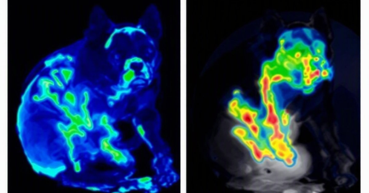
What the Researchers Did
This study took a focused, practical approach. The team analyzed CT scans from 10 American Staffordshire Bull Terriers — all healthy dogs with no shoulder problems. Using the CT images, evaluators traced the outlines of two specific shoulder muscles:
- The supraspinatus — the muscle that sits on top of the shoulder blade and helps lift the leg forward
- The infraspinatus — the muscle at the back of the shoulder blade, important for rotating and stabilizing the joint
This tracing process is called segmentation — essentially drawing a careful outline around each muscle on the scan, slice by slice, so a computer can calculate its total volume (size). Think of it like using cookie cutters on stacked slices of bread: you trace the shape on each slice and then add up all those shapes to get the full 3D volume.
The same scans were then reviewed multiple times, by the same evaluator on different days and by different evaluators, to test how consistent the measurements were.
What the Study Found
Near-Perfect Reliability Across the Board
The standout finding was how consistently the measurements lined up. When the same evaluator measured the same scan on different days, the scores were nearly identical. When two different evaluators measured the same scan independently, they still reached the same conclusions. The reliability scores — called intraclass correlation coefficients (a statistical measure of agreement, where 1.0 means perfect) — ranged from 0.958 to 0.996.
To put that in everyday terms: if two vets each measured a dog’s shoulder muscle using this method, their results would differ by less than 5% — which is well within the margin of precision needed to confidently track changes over time.
A Repeatable Tool for Monitoring Recovery
The researchers concluded that manual CT segmentation is a reliable method for measuring shoulder muscle volume in dogs. That means the same measurement approach, used weeks apart or by different clinicians, will produce results consistent enough to actually trust when comparing one visit to the next.
For rehabilitation — where the whole goal is to see whether muscles are recovering — that consistency is everything. Without it, you can’t tell whether a muscle is growing back, staying the same, or continuing to shrink.
What This Means for Your Dog
Smarter Monitoring During Rehab
If your dog is recovering from a shoulder injury, surgery, or orthopedic condition affecting the front legs, this type of imaging-based tracking could eventually become part of a more data-driven rehab plan. Instead of relying entirely on how your dog looks or moves, your vet or rehabilitation specialist could potentially use CT measurements at different points in the recovery process to confirm that muscle mass is actually coming back.
This is particularly useful for dogs that are stoic — many dogs mask pain or discomfort well, making it hard to tell from behavior alone how recovery is progressing.
What You Can Do Right Now
While this technology isn’t yet standard practice at every clinic, there are practical steps you can take if your dog has a shoulder problem:
- Ask about specialist referrals — Veterinary rehabilitation specialists and orthopedic vets are more likely to use advanced imaging tools and have access to CT scanning.
- Ask about baseline imaging — If your dog is having shoulder surgery or starting a rehab program, ask whether a pre-treatment CT scan might be useful as a baseline to compare against later.
- Track what you can at home — Note your dog’s activity level, willingness to bear weight on the front leg, and any visible differences in shoulder shape. Bring those observations to every vet visit.
When to Talk to Your Veterinarian
Reach out to your vet if your dog:
- Limps on a front leg or favors one side consistently
- Seems reluctant to run, jump, or climb stairs as usual
- Flinches or pulls away when the shoulder area is touched
- Has visible muscle differences between the two shoulders
- Is recovering from a shoulder injury but doesn’t seem to be improving as expected
Early assessment means earlier intervention — and the sooner muscle atrophy is caught, the better the chances of a full recovery.
Study Limitations to Keep in Mind
This was a small study with just 10 dogs, all of whom were healthy. The reliability findings are promising, but they don’t yet tell us how the method performs in dogs that are already injured or sick, where swelling, scarring, or changes in tissue density might make the tracing process harder. The study also measured the muscle segmentation technique itself — not whether using it actually leads to better outcomes for patients.
Larger studies across different breeds, ages, and injury types will be needed before CT-based muscle measurement becomes a standard tool in everyday vet clinics. But as a proof-of-concept showing the method is consistent and repeatable, this study lays solid groundwork.
The Bottom Line
Reliably measuring dog shoulder muscle loss has long been a challenge in veterinary medicine. This study shows that CT scans, combined with careful muscle tracing, can produce highly consistent results — with reliability scores of 0.958 to 0.996. That kind of precision could transform how vets monitor shoulder injuries and track rehabilitation progress, replacing guesswork with data.
If your dog has had a shoulder problem or is heading into surgery, it’s worth having a conversation with your vet about imaging options and what rehab monitoring might look like. The science is pointing toward a future where tracking your dog’s muscle recovery is as measurable and reliable as checking any other vital sign.
This article summarizes peer-reviewed research for educational purposes. Always consult with your veterinarian for personalized advice about your pet’s health and behavior.