A Smarter Way to See Inside a Dog’s Liver Tumor
Dog liver tumor treatment just got more precise: a new study found that a special type of 3D imaging used during the procedure can reveal hidden blood vessels feeding the tumor — vessels that a standard CT scan missed entirely in 5 out of 36 dogs. Catching those vessels matters a lot, because if you miss them, you miss part of the tumor’s blood supply — and the treatment may not work as well.
Published in Frontiers in Veterinary Science, this research looked at a specific liver cancer treatment called transarterial chemoembolization — more on that below. The key takeaway for pet owners: better real-time imaging during this procedure could mean more thorough treatment and less exposure to radiation and contrast dye for your dog.
Why Liver Tumors in Dogs Are So Hard to Treat
The liver has a rich blood supply, which is one reason it’s a common site for tumors in dogs. Liver tumors get most of their nutrients and oxygen from blood vessels that feed directly into them. One of the most effective ways to treat these tumors is to target those very blood vessels — cutting off the tumor’s “food supply” while delivering chemotherapy right to the source.
The procedure used in this study is called hepatic transarterial chemoembolization, or TACE for short. Think of it like a precision delivery system: a tiny tube (called a catheter) is guided through the blood vessels into the ones feeding the tumor. Then doctors inject a mixture of chemotherapy drugs and a substance that blocks the blood flow. The tumor is essentially starved and dosed at the same time, while the rest of the liver and body are largely spared.
The challenge? To do this accurately, veterinarians need to see exactly which blood vessels are feeding the tumor. If they miss one, the tumor can keep getting a blood supply — and the treatment loses effectiveness.
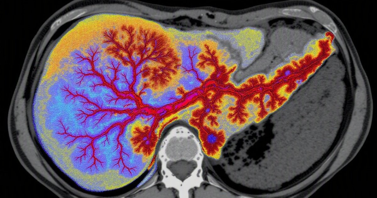
What Is Cone-Beam CT — and How Is It Different?
Before this study, vets relied on a regular CT scan done before the procedure to map out the tumor’s blood vessels. That works most of the time. But CT scans are taken in the imaging suite, not in the operating room. By the time the procedure begins, the vet is working from a “map” made earlier — and some vessels may not have been visible.
The newer tool used in this study is called cone-beam CT, or CBCT. It’s a type of 3D X-ray machine that can be used right there in the procedure room, while everything is happening. Instead of a single flat image, it creates a detailed three-dimensional picture of the blood vessels around the tumor in real time. Think of the difference between a regular city road map and a live GPS with traffic updates — one is a snapshot from earlier, the other shows you exactly what’s happening right now.
How the Study Was Done
Researchers looked back at records from 36 client-owned dogs — real pets, not lab animals — who had all undergone TACE for liver tumors. The dogs were split into two groups of 18:
- Group 1 had their procedure guided by standard preoperative CT — the traditional approach, using scans taken before the procedure
- Group 2 had their procedure guided by intraoperative cone-beam CT — the newer approach, with real-time 3D imaging during the procedure
Because this was a retrospective study (meaning researchers looked back at past cases rather than running a new experiment), they were able to compare what was found and how much radiation and contrast dye (the substance injected to make blood vessels visible on scans) each group was exposed to.
What the Study Found
Hidden Vessels That Standard CT Missed
The most striking finding: in 5 of the 18 dogs in the cone-beam CT group, the real-time imaging revealed tumor-feeding blood vessels that had not been visible on the standard preoperative CT scan.
These weren’t small or unimportant vessels. They were extrahepatic (outside the liver itself) vessels still feeding the tumor. If those vessels had gone undetected, part of the tumor’s blood supply would have been left untouched — potentially allowing the tumor to keep growing.
This means that roughly 1 in 4 dogs in the cone-beam CT group had important vessels that would have been missed using the older method alone. That’s a significant gap in the information vets were working with.
Less Radiation and Less Contrast Dye
The study also found that dogs in the cone-beam CT group were exposed to lower doses of radiation and required less contrast medium (the dye used to make blood vessels show up on imaging). This might seem counterintuitive — you might expect a more advanced imaging technique to use more radiation — but cone-beam CT is specifically designed to give detailed pictures with less exposure than traditional methods.
For a dog undergoing treatment for a serious condition, reducing unnecessary radiation and dye is a real quality-of-life and safety benefit.
What This Means for Dog Owners
More Complete Treatment May Be Possible
If your dog has a liver tumor and your vet is considering a minimally invasive treatment like TACE, this study suggests that access to intraoperative cone-beam CT could make the procedure more thorough. By seeing the full picture of blood vessels in real time, the team is less likely to miss a vessel that could undermine treatment.
This kind of technology isn’t available everywhere. It tends to be found at veterinary specialty centers or academic veterinary hospitals with advanced imaging equipment. If your dog is being treated for liver cancer, it’s worth asking whether your treatment facility has this capability — or whether a referral to a specialty center makes sense.
When to Consult Your Veterinarian
Talk to your vet or a veterinary oncologist if:
- Your dog has been diagnosed with a liver tumor and you’re discussing treatment options
- You’ve been referred for a procedure like TACE and want to understand what imaging will be used
- You want to know whether a specialty center with advanced imaging technology is nearby
- Your dog has a known history of liver disease and you’re monitoring for new growths
Your vet can help you understand what facilities and techniques are available in your area and whether your dog is a good candidate for minimally invasive liver tumor treatment.
Study Limitations
This was a retrospective study — meaning researchers looked back at existing records rather than designing a controlled experiment from the start. That approach can introduce selection bias, where the dogs in each group may have differed in ways that influenced the results (for example, if one group tended to have more complex cases). The sample size of 36 dogs is also relatively small, so the findings — while promising — need to be confirmed in larger, prospective trials. We also don’t have long-term follow-up data on how these dogs did after treatment. Future studies should track whether better vessel detection actually leads to longer survival or slower tumor progression.
The Bottom Line
A study of 36 dogs found that real-time 3D cone-beam CT imaging during liver tumor treatment detected hidden tumor-feeding blood vessels in 5 cases that a standard pre-procedure CT scan had missed. It also reduced the radiation dose and contrast dye needed. For dogs with liver tumors undergoing minimally invasive treatment, access to this kind of advanced imaging could mean a more complete procedure and a safer experience overall.
This technology isn’t yet available everywhere, but it’s an important step forward in veterinary oncology. If your dog is facing liver tumor treatment, it’s worth asking your vet whether intraoperative imaging options are available — or whether a referral to a specialty center makes sense.
This article summarizes peer-reviewed research for educational purposes. Always consult with your veterinarian for personalized advice about your pet’s health and behavior.