Pre-Surgery Scans Are Highly Reliable for Pet GI Problems
When your dog or cat needs surgery for a stomach or intestinal problem, the scans your vet takes beforehand are right about 88% of the time. That’s the key finding from a new study published in Frontiers in Veterinary Science, which compared pre-surgery imaging results to what surgeons actually found when they operated on 95 pets.
For pet owners, this is reassuring news. It means that when your vet looks at a scan and tells you what’s going on inside your pet’s belly, they’re most likely correct—and that the surgery plan is built on solid information.
Why Imaging Before Surgery Matters
When a pet has serious stomach or intestinal trouble—think vomiting, bloating, or signs of a blockage—surgery is sometimes the only answer. But before a vet makes the first incision, they need to know what they’re dealing with. That’s where pre-surgery imaging comes in.
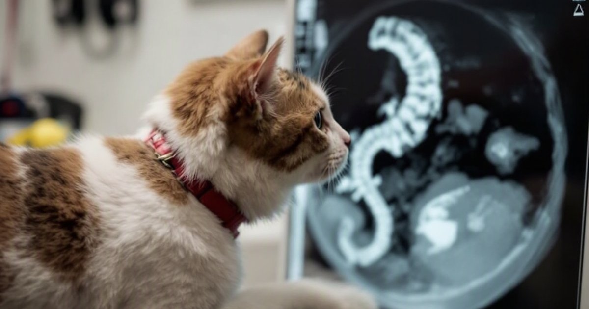
Vets use two main tools to see inside a pet’s abdomen (belly):
- X-rays (radiography) — a quick, widely available image that shows bones and organs
- Ultrasound — a scan that uses sound waves to create a moving picture of soft tissues, similar to what you see when a doctor checks a baby before birth
The question researchers wanted to answer: How often do these scans get it right? Knowing the answer helps vets and pet owners trust the information they’re basing major decisions on.
About the Study
This research looked back at the medical records of 95 pets—35 cats and 60 dogs—who had all come to a vet clinic showing signs of gastrointestinal (GI) trouble, meaning problems with their stomach or intestines. All of these animals went on to have an exploratory surgery called a laparotomy (a procedure where the surgeon opens the belly to get a direct look inside).
The researchers then compared two things side by side:
- What the pre-surgery scans (X-rays and ultrasound) had suggested was wrong
- What the surgeon actually found once inside
This type of study—called a retrospective study—means it looked back at cases that had already happened, rather than setting up a new experiment from scratch.
What the Researchers Found
Imaging and Surgery Agreed Most of the Time
In about 88% of cases, what the scans showed before surgery matched what the surgeon found when they opened the pet up. That’s a strong level of agreement. It tells us that vets can generally trust their imaging tools when planning surgery for GI issues in dogs and cats.
X-Rays Outperformed Ultrasound
One of the study’s more notable findings: for identifying the main problem in the GI tract, X-rays did a better job than ultrasound. This might seem surprising, since ultrasound is often seen as the more high-tech option. But for certain GI issues—like a blockage or a mass—X-rays can be more straightforward and effective at pinpointing the culprit.
What This Means for You and Your Pet
You Can Trust Your Vet’s Pre-Surgery Plan
If your vet tells you that imaging shows your dog has an intestinal blockage or your cat has a suspicious mass in their belly, this research backs up that assessment. The scans vets use are not perfect, but they’re right the vast majority of the time—and they directly shape how a surgery is planned.
X-Rays Are a Valuable First Step
Many pet owners assume ultrasound is always the “better” option because it’s more advanced. This study suggests that for GI issues specifically, X-rays shouldn’t be overlooked. They may actually give the vet a clearer picture of the main problem.
When to Consult Your Veterinarian
If your pet is showing signs of GI trouble—repeated vomiting, a swollen belly, not eating, pain when the belly is touched, or straining to go to the bathroom—it’s important to see a vet promptly. These can be signs of conditions that require imaging and, in some cases, surgery. Don’t wait to see if it gets better on its own.
Study Limitations
Like all research, this study has some boundaries. It was a retrospective study, meaning it analyzed past cases rather than running a controlled experiment. The findings are based on a specific group of 95 pets at one or a few clinics, so results might look different in other settings or with different types of GI conditions. The study itself notes that the conclusions may not apply to every kind of gastrointestinal case. More research with larger, more varied groups of pets would help confirm and expand these findings.
The Bottom Line
Pre-surgery imaging is a powerful and reliable tool when your dog or cat faces gastrointestinal surgery. This study found that X-rays and ultrasound scans correctly predicted what surgeons would find about 88% of the time across 95 pets—and that X-rays were especially strong at identifying the main problem.
If your vet recommends imaging before a GI procedure, this research supports that approach. Accurate scans help surgeons go in prepared, which can make a real difference in outcomes for your pet.
This article summarizes peer-reviewed research for educational purposes. Always consult with your veterinarian for personalized advice about your pet’s health and behavior.